


wangbz 提交于
报道:来自瑞典卡罗林斯卡医学院著名的发育生物学家Kaj Fried和Igor Adameyko发现,牙齿软组织内的间充质干细胞有一个意想不到的来源,即外周神经胶质细胞。研究结果发表在最新的Nature杂志上,将有助于揭示牙齿如何形成,如何成长,如何能自我修复。
2014年7月27日 《自然》
![]()
中文翻译
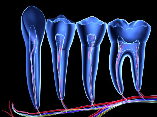
【题目】 牙齿模型系统中发现来源于神经的间充质干细胞
【译文】 间充质干细胞聚集在基质组织中,在细胞生长和修复过程中提供特定的间充质衍生物。间充质干细胞的来源是被热烈讨论的话题之一,目前一致认为血管周围细胞是形成间充质干细胞的最佳组织。不断增长的小鼠门齿为我们提供了一个很好的模型来解决间充质干细胞的来源。这些干细胞存在于能产生各种特异性衍生物的牙根尖。构成牙齿的细胞主要有两个胚胎来源:神经嵴的外胚层间质和外胚层上皮细胞。这几十年来一直认为牙间充质干细胞能引起牙髓细胞和成牙本质细胞(来源于神经嵴细胞)在早期脑部的迁移以及间充质组织的形成。在本文中,我们的结果表明,在牙齿发育期间,相当数量的能自我更新和修复牙齿的间充质干细胞来自于外周神经胶质细胞。神经胶质细胞可以转化为多功能间充质干细胞,而这些干细胞可以发育成牙髓细胞和成牙本质细胞。通过结合彩色编码克隆技术追踪外周神经胶质细胞,我们在牙齿器官生成和生长方面提供了新的动力学见解。
英文原稿
[Title] Glial origin of mesenchymal stem cells in a tooth model system
[Author] Nina Kaukua, Maryam Khatibi Shahidi, Chrysoula Konstantinidou, Vyacheslav Dyachuk, Marketa Kaucka, Alessandro Furlan, Zhengwen An,
Longlong Wang, Isabell Hultman, Lars A ¨hrlund-Richter, Hans Blom, Hjalmar Brismar, Natalia Assaife Lopes, Vassilis Pachnis, Ueli Suter,
Hans Clevers, Irma Thesleff, Paul Sharpe, Patrik Ernfors, Kaj Fried & Igor Adameyko
[Abstract] Mesenchymal stem cells occupy niches in stromal tissues where they provide sources of cells for specialized mesenchymal derivatives during
growth and repair. The origins of mesenchymal stem cells have been the subject of considerable discussion, and current consensus holds that
perivascular cells form mesenchymal stem cells in most tissues. The continuously growing mouse incisor tooth offers an excellent model to
address the origin of mesenchymal stem cells. These stem cells dwell in a niche at the tooth apex where they produce a variety of differentiated
derivatives. Cells constituting the tooth are mostly derived from two embryonic sources: neural crest ectomesenchyme and ectodermal epithelium.
It has been thought for decades that the dental mesenchymal stem cells giving rise to pulp cells and odontoblasts derive from neural crest cells
after their migration in the early head and formation of ectomesenchymal tissue. Here we show that a significant population of mesenchymal stem
cells during development, self-renewal and repair of a tooth are derived from peripheral nerve-associated glia. Glial cells generate multipotent
mesenchymal stem cells that produce pulp cells and odontoblasts. By combining a clonal colour-coding technique with tracing of peripheral glia,
we provide new insights into the dynamics of tooth organogenesis and growth.
原文地址: http://www.nature.com/nature/journal/vaop/ncurrent/full/nature13536.html
- 浏览 6544 次

